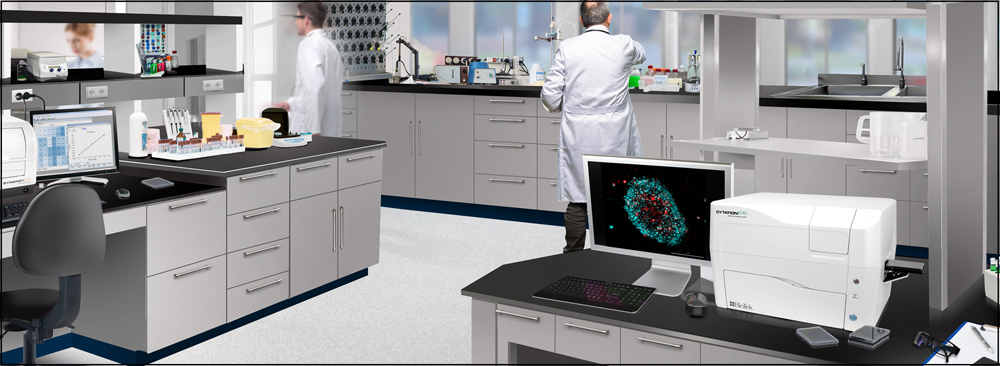
Cytation™ C10 Confocal Imaging Reader combines automated digital confocal and widefield microscopy with conventional multi-mode microplate reading in a unique, patented design. The spinning disk confocal module provides exquisite resolution and optical sectioning capabilities in a wide variety of sample types. High quality components used in the system, including a Hamamatsu scientific CMOS (sCMOS) camera, Olympus objectives and laser-based illumination enable excellent image quality, at a significantly lower price than others. Cytation C10 also includes widefield fluorescence, brightfield and phase contrast optics. Its variable bandwidth monochromator-based multi-mode plate reading is based on the proven design of the market-leading BioTek Synergy™ products. Environmental controls enable live cell assays, and Gen5™ software is specifically designed to make sample detection and image capture quick and effortless. The new 3D viewer provides users with 3D reconstruction of their thicker samples captured with the confocal microscope. Cytation C10 brings affordable confocal to every laboratory.
◆ 3D cell imaging and analysis
◆ Cell migration and invasion assays
◆ Live cell imaging
◆ Cell proliferation
◆ Cell viability/toxicity
◆ Immunofluorescence
◆ Label-free cell counting
Detailed Characterization of Mitosis Using Cell Population Analysis and Auto ROI Defined Confocal Imaging
Confocal Imaging of Spheroids for Accurate Determination of Cell Number and Evaluating Treatment Response
特色
Compact, affordable confocal imager for every laboratory
Expertise gained over several years of Cytation development, along with customer feedback, resulted in the Cytation C10.... an automated confocal microscope with excellent performance at a truly attainable price.
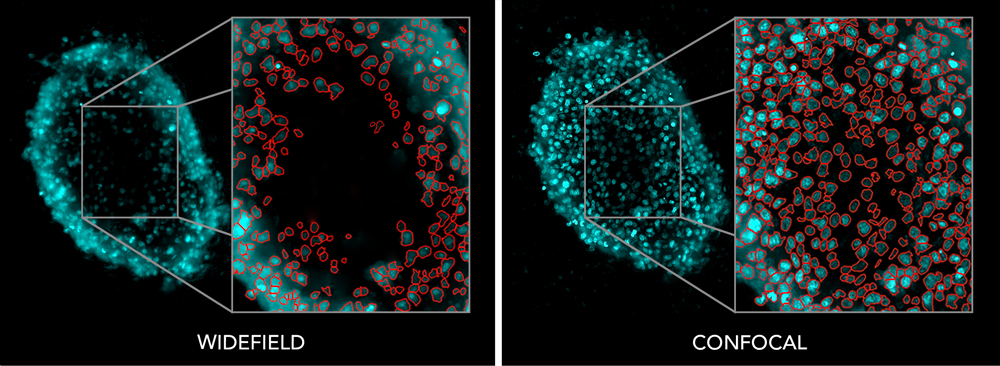
Confocal: Improved image quality and analysis
Confocal microscopy can enable you to see a level of detail in your samples that is not possible with widefield optics. Not only can you obtain improved image quality, you can get improved quantification and analysis with confocal images and Gen5 software.
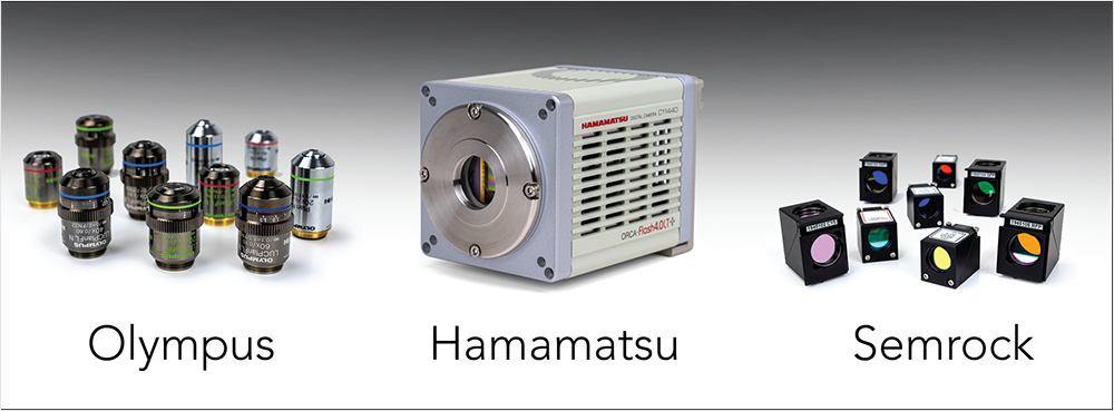
High quality optical components
High quality objectives, filters and other components including the Hamamatsu sCMOS Orca camera, Semrock filters, Olympus objectives and other well-known brands, are used in Cytation C10, enabling the capture of stunning, publication-quality images.
Confocal imaging and multi-mode plate reader in one
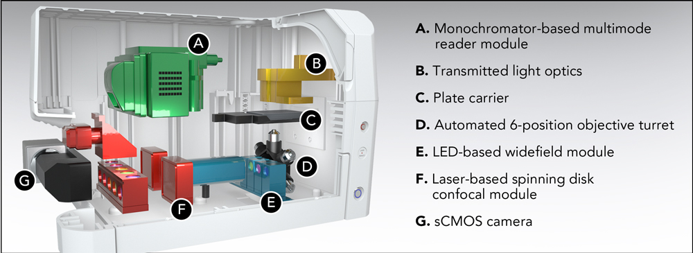
With a combination of spinning disk confocal and widefield imaging, plus multi-mode reader, Cytation C10 is truly ready for any assay. And since Cytation C10 is a modular, upgradable instrument, you can get the functionality you need today and add modules later as your needs expand.
Confocal plus widefield = stunning images and analysis
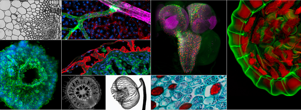
Cytation C10 captures stunning detail in a wide variety of sample types. Use widefield imaging for faster acquisition of large samples at lower magnification, switch to confocal to image small intracellular details or 3D samples. Or combine both modes for highly multiplexed, multi-parameter imaging experiments.
Environmental controls for live cell imaging
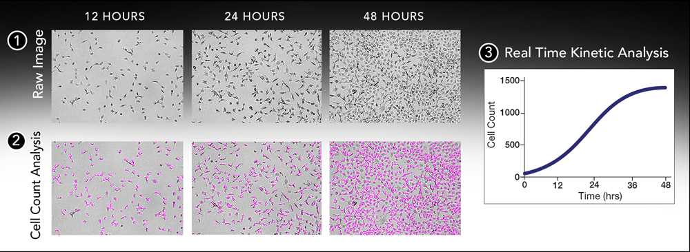
Successful live cell kinetic imaging relies on a consistent environment, including temperature control and CO2/O2 control and monitoring. Cytation C10 provides the perfect environment to grow and analyze live cells over time. Powerful movie maker and kinetic analysis software tools allow visualizing and analysis time-lapse experiments.
Hit-picking: Multi-mode detection + imaging saves time and data storage
(1) Plate reader quickly identifies GFP positive wells.
(2) Only GFP positive wells are imaged, saving both time and computer memory.
Imaging data sets can take a long time to acquire and require large data storage capacity. The unique hit-picking function takes advantage of the embedded plate reader optics. Set the hit picking criteria, quickly pre-screen the microplate with the plate reader optics and Cytation C10 will automatically image the samples that meet your criteria, saving both time and hard drive space.
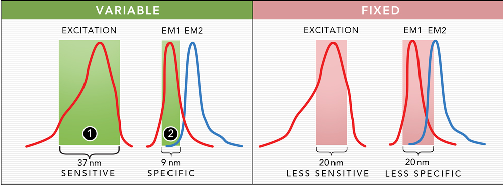
Variable bandwidth for sensitivity and specificity
The plate reader optics of Cytation use a quad monochromator design with variable bandwidth. The bandwidth can be set anywhere between 9 and 50 nm in 1 nm increment. Large bandwidth settings (1) provide increased sensitivity and lower limits of detection. Small bandwidth settings (2) provide increased specificity when multiple signals are present, which reduces signal crosstalk and enhances assay performance.
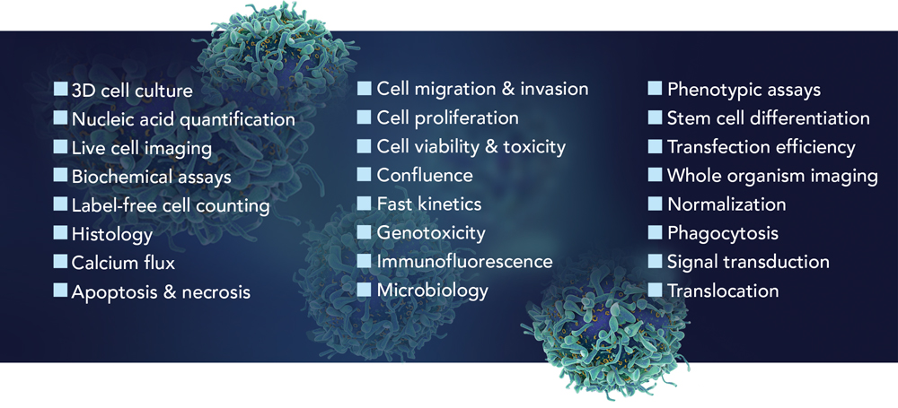
Cytation C10: Ready for any application
The combination of confocal and widefield imaging with multi-mode detection will transform your laboratory workflows and increase productivity. Cytation C10 is truly ready for any application.
For Research Use Only. Not for use in diagnostic procedures.
Endpoint, kinetic, spectral scanning, well area scanning
Microplate types
Monochromator: 6- to 384-well plates
Imaging: 6- to 1536-well plates
Other labware supported
Microscope slides, Petri and cell culture dishes, cell culture flasks (T25), counting chambers (hemocytometer), Take3 Micro-volume plates
Temperature control
4-Zone incubation to 45 °C with Condensation Control
Shaking
Linear, orbital, double-orbital
Software
Gen5 Microplate Reader and Imager Software included
Gen5 Secure for 21 CFR Part 11 compliance (option)
Gen5 Image+ and Image Prime software available for full image analysis (option)
Automation
BioStack and 3rd party automation compatible
CO2 and O2 control (option)
Range: 0 - 20% (CO2); 1 - 19% (O2), with optional Gas Controller
Models for both CO2 and O2 or CO2 only are available
IMAGING - CONFOCAL MICROSCOPE
Imaging modes
Fluorescence
Image processing
Z-projection, digital phase contrast, stitching
Camera
Hamamatsu Orca sCMOS, 16-bit grayscale camera or
Sony CMOS 16-bit grayscale camera
Objective capacity
6-position automated turret for user-replaceable objectives